
Contact Us

Silent MRI
Silent MRI Scan is a promising technique for acoustic noise reduction and improved patient comfort.
3T Platform
Silent MRI
Silent MRI Scan is a promising technique for acoustic noise reduction and improved patient comfort.
- 97% less noise
- 100% more patient-friendly
- reasonable noise reduction
- Latest and advanced MRI technology
- Superior results and image quality

CT Scan
CT Scan uses a combination of X-rays and a computer to create pictures of your organs, bones, and other tissues
True 16 detectors- 96 Slice Reconstruction
CT Scan
CT Scan uses a combination of X-rays and a computer to create pictures of your organs, bones, and other tissues
- More detailed than a regular X-ray
- Painless, non-invasive, and accurate
- Fast & simple
- No radiation remains in a patient's body

Digital X-ray
Digital X-ray is a form of X-ray imaging, where digital X-ray sensors are used.
DDR - 500 mA
Digital X-ray
Digital X-ray is a form of X-ray imaging, where digital X-ray sensors are used.
- Time efficiency through bypassing chemical processing
- Ability to digitally transfer and enhance images
- Faster processing time
- Allows for higher quality care

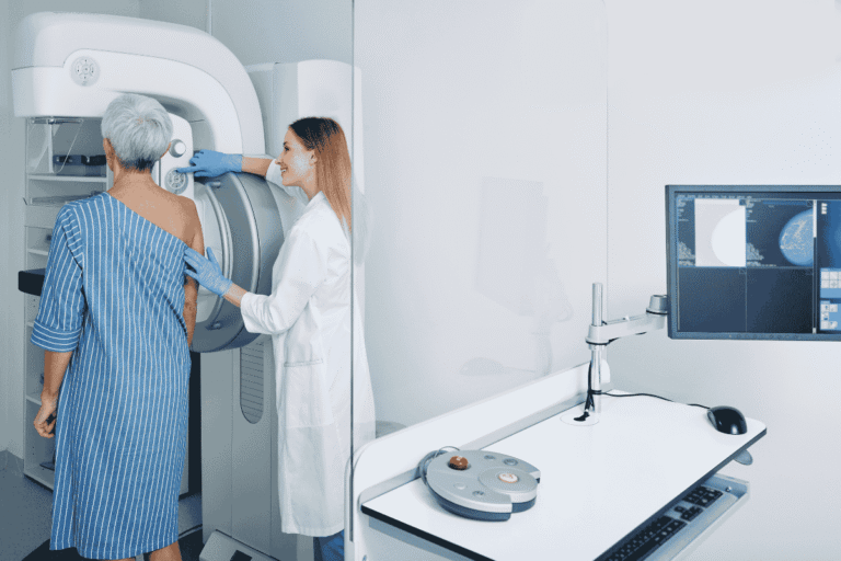
Digital Mammography
Digital mammography is a mammography system in which the X-ray film is replaced by solid-state detectors that convert X-rays into electrical signals
Digital Mammography
Digital mammography is a mammography system in which the X-ray film is replaced by solid-state detectors that convert X-rays into electrical signals
- Quick availability of results
- 25% less radiation than traditional methods
- Enhances patient care with precision
- Uses solid-state detectors for clarity

3D/4D Ultrasound with Color Doppler
Ultrasound examinations help to diagnose a variety of conditions and to estimate organ damage following any illness. Ultrasound produces pictures of the inside of the body using sound waves
3D/4D Ultrasound with Color Doppler
Ultrasound examinations help to diagnose a variety of conditions and to estimate organ damage following any illness. Ultrasound produces pictures of the inside of the body using sound waves
- Completely safe and painless
- Real-time visualization of the fetus or organs
- Non-invasive
- Helps to identify structural inherent anomalies of the fetus during the scheduled 18-20 week scan.

Barium
It is a study for the gastrointestinal tract. It is generally used to highlight abnormalities in the GI tract.
Special X-ray Investigations
Barium
It is a study for the gastrointestinal tract. It is generally used to highlight abnormalities in the GI tract.
- Helps determine the cause of painful swallowing, abdominal pain, blood-stained vomit, or unexplained weight loss.

HSG
It studies the inside of the uterus and fallopian tubes and the area around them. It is often done in women who are infertile and having difficulty getting pregnant.
Special X-ray Investigations
HSG
It studies the inside of the uterus and fallopian tubes and the area around them. It is often done in women who are infertile and having difficulty getting pregnant.
- Allows your doctor to more accurately investigate the cervical areas
- Increases pregnancy successes

IVP
This is an X-ray test that provides images of the kidneys, the urinary bladder, the ureters, and the urethra (urinary tract). An IVP can show the size, shape, and position of the urinary tract, and…
Special X-ray Investigations
IVP
This is an X-ray test that provides images of the kidneys, the urinary bladder, the ureters, and the urethra (urinary tract). An IVP can show the size, shape, and position of the urinary tract, and…
- Provides enough information about kidney stones and urinary tract obstructions to treat with medication and avoid invasive surgical procedures
- No radiation remains in a patient's body after an x-ray examination

Fistulogram
Precise fistula assessment with advanced technology for patient care
Special X-ray Investigations
Fistulogram
Precise fistula assessment with advanced technology for patient care
- Assesses fistula conditions accurately
- Guides treatment with real-time imaging
- Enhances patient care planning
- Non-invasive procedure


Dexa
DEXA uses a very small dose of ionizing radiation to produce pictures of the inside of the body (usually the lower spine and hips) to measure bone loss.
BMD
Dexa
DEXA uses a very small dose of ionizing radiation to produce pictures of the inside of the body (usually the lower spine and hips) to measure bone loss.
- Can diagnose osteoporosis with ease
- Assesses an individual's risk for developing osteoporotic fractures
- Prevents disabilities and fractures
- Analyzes the calcium content and density of bones

Interventions USG/CT guided
Ultrasound (US) is often used to guide various interventional procedures in the genitourinary (GU) tract because it can provide real-time imaging without any radiation hazard.
Interventions USG/CT guided
Ultrasound (US) is often used to guide various interventional procedures in the genitourinary (GU) tract because it can provide real-time imaging without any radiation hazard.
- Real-time imaging without radiation
- Guides genitourinary procedures safely
- Enhances patient care with precision
- Non-invasive technique

Fetal Echocardiography
A test similar to an ultrasound, Fetal Echocardiography allows doctors to examine the structure and function of your unborn child’s heart. It’s typically done in the second trimester, between weeks 18 to 24.
Fetal Echocardiography
A test similar to an ultrasound, Fetal Echocardiography allows doctors to examine the structure and function of your unborn child’s heart. It’s typically done in the second trimester, between weeks 18 to 24.
- Prenatal diagnosis of congenital heart disease (CHD)
- Evaluates the position, size, structure, function, and rhythm of the unborn baby’s heart.

Fibroscan
Non-invasive liver assessment with advanced technology for accuracy.
- Measures liver stiffness and fat non-invasively
- Detects fibrosis or fatty liver disease
- Uses advanced technology for precise results
- Quick and comfortable procedure
HRCT Chest
High-resolution COVID scan with advanced technology.
- High-resolution COVID scan.
- Detailed imaging with advanced technology.
- Non-invasive and fast.
- Ensures accurate reports.